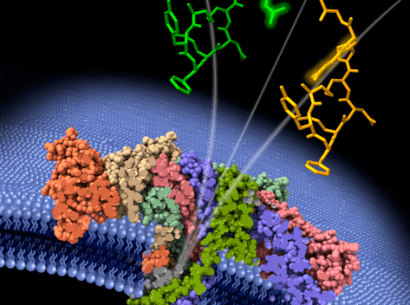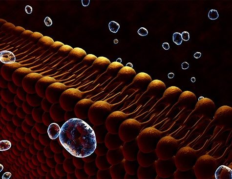
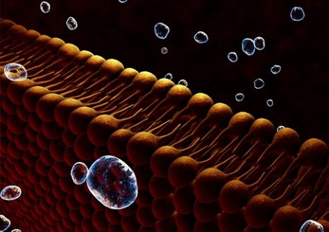
The cell membrane is a constantly-evolving structure. Under the influence of the cytoskeleton, (filaments that ensure cell rigidity and internal molecular movement), it can change its shape greatly, even forming cylinders in its surface. These cylinders, called nanotubes, appear and disappear in a dynamic manner to bring biological molecules into the cell.
Atomic force microscopy to study the cell membrane surface
Lambe’s Interface Polymer Materials team (University of Évry /CNRS/CEA/Cergy-Pontoise University) developed an experimental platform to elucidate nanotube mechanics at the nanometric scale. Their approach employs atomic force microscopy (AFM), a technology acquired by the lab and falling within the competencies of its researchers. The work, carried out in partnership with the Institut Curie and the Laboratory of Theoretical Physics and Statistical Models (LPTMS; Paris-Saclay University), was published in Physical Review X in February of 2020.
AFM uses a type of cantilever-mounted probe, the nanometric tip of which is placed in contact with the cellular membrane. There, it sounds the surface, measuring the elasticity and topographic changes occurring as it moves. In that way, the researchers gathered information on the morphology, size and rigidity of the nanotubes, thus gaining insights into their mechanics. Their initial study thus revealed information on the relationship between nanotube size and rigidity, and on their behavior under lateral compression. The method deployed by Lambe opens new frontiers of study. Particularly, it will contribute to knowledge on the role of cellular proteins on nanotube remodeling.
With its experimental platform, at the crossroads of physics and biology, Lambe is helping to dissect the physical mechanisms underlying fundamental processes in intracellular trafficking.